


Osteochondrosis of the lumbar region is a chronic degenerative dystrophic disease of the lumbar spine that affects the structure of the intervertebral discs and the lumbar spine.This affects mainly people of working age.It manifests itself in various symptoms, the main thing that is pain in the lower back and legs, lower back movements.Research methods such as radiography, computed tomography, or lumbar spine magnetic -resonance imaging are used for diagnosis.In this article you can detail in more detail, the causes, symptoms and methods of diagnosing lumbar osteochondrosis.
Osteochondrosis is the result of aging of the body.You can see these or other signs of this disease in almost every person (!), It starts at the age of 25.But here is the severity of these changes, the speed of their progression, the quality of clinical manifestations depends on many reasons, primarily on how a healthy lifestyle leads to a particular person.Moderate physical activity, mandatory morning gymnastics, right posture of the body, when performing numerous work (garden, construction, banal cleaning of the house, etc.), orthopedic mattresses are moments that prevent lumbar spine osteochondrosis.
According to statistics, osteochondrosis of the spine in 80% of cases is the cause of spinal pain.
How does osteochondrosis develop?
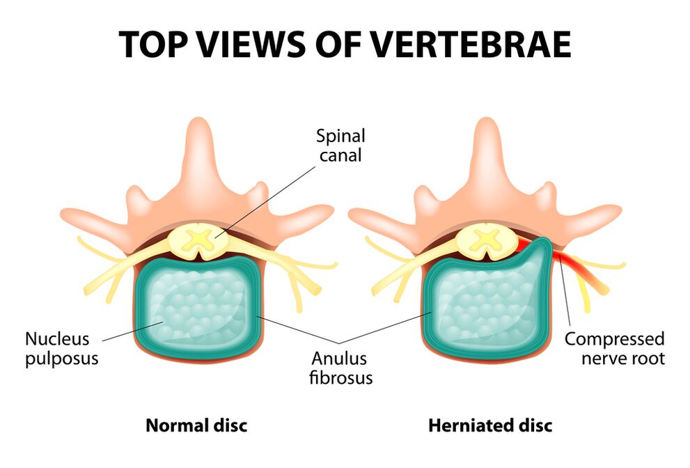
The whole spine contains separate vertebrates, between the bodies of which there are intervertebral discs.That is, there is one disc between the two vertebrates.The disc is made up of a gelatin (pulpic) nucleus and a fibrous rim.The nucleus contains a lot of water and provides spine depreciation and flexibility.The fiber ring is located along the periphery of the jacket nucleus, as if holding it inside.
With a prolonged load on the dirt nucleus, it alters its physiological properties, loses water and dries, and eventually in sequence: the disc is flattened, and the spine bodies approach.In addition to such processes, in the nucleus jacket, the fiber link loses its elasticity and, under the influence of mechanical loads, begins to be clutched.This is called protrusion.Then the fiber ring cracks and the gelatin nucleus passes through the resulting gaps: the disk hernia becomes.The plot of two adjacent spine, including the disk, which is called the spine segment, gains excessive mobility, thereby increasing the load on nearby segments.Overloading of neighboring segments leads to a similar pathological process in them.These changes are called osteochondrosis.
To some extent ensuring the stability of the spine, the growth of the bones arises along the edges of the spine organs, increasing the support area.This phenomenon is called spondylosis.Changes in the joints between the vertebrates are called spondylo arthrosis.Usually all three pathologies - osteochondrosis, spondylosis, spondil arthrosis - go there.
Causes
Why Osteochondrosis?To date, there are several theories of events:
- Mechanical theory: Perhaps the main cause should be considered a regular load of the spine.That is why osteochondrosis is an almost mandatory fate of movement, miners, builders, and people of such professions.Osteochondrosis of the lumbar region is mainly associated with slopes and gravity, forcing uncomfortable working posture;
- Another factor in development is the wrong posture sitting in the wrong posture, which is especially relevant for mental workers;
- Sometimes the hereditary properties of the spine structure and the nutrition of its individual structures play a role;
- Traumatic theory: Any trauma to the spine (the most minor) can lead to a degenerative process;
- Hormonal metabolic disorders and endocrine diseases can adversely affect metabolism in the spinal column tissues and promote the development of osteochondrosis;
- Age theory involves the natural wear of discs in the process of living.
Rarely, only one of these theories can explain osteochondrosis in each case.More often at once, several factors are "guilty".
In the case of lumbar spine osteochondrosis, excess weight plays an important role as it is in itself overloading the spinal column.The higher the body mass index (the degree of obesity), the more pronounced changes in the spine usually occur.Other causes that provoke osteochondrosis may be:
- Sedentary lifestyle;
- Imorly meals (fast food, excessive sweet, semi -coated products: all this causes trace elements imbalances) and lack of fluid;
- Anomalies of the spine structure (for example, the presence of additional lumbar spine);
- Constant wear with high shoe shoes;
- Pregnancy (due to excessive lumbar spine load);
- Sudden discontinuation of training in people professionally involved in sports;
- Alcohol smoking and abuse: As a factors that accelerate the aging process in the body.
Symptoms
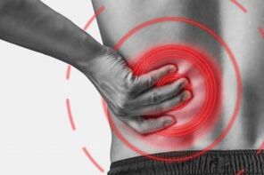
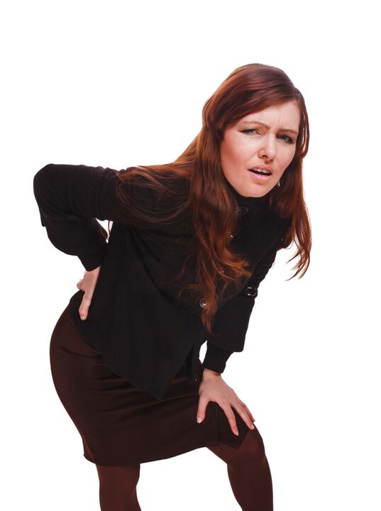
The main manifestation of lumbar spine osteochondrosis is pain.The nature of the pain, the scene of the accident, and the direction of distribution depends on which receptors are irritated, that is, how rough changes are the disk and the surrounding tissues, there is protrusion or hernia in which direction protrusion, etc.
Reflex and compression syndromes are characterized by osteochondrosis of the lumbar spine.
Reflex syndromes develop in cases where the affected disk fiber -ring receptors, ligaments and nearby joint capsules are irritable.They are reflexive because in addition to pain, it is accompanied by muscular, vegetative-vascular or neurodystrophic reflex changes, that is, irritation with reflexes transmits to other structures, causing symptoms mainly on soft tissues.
Compression syndromes occur as a result of nerve roots, blood vessels, or spinal cord compression (compression) produced by osteochondrosis, changes.

Reflex syndromes of lumbar spine
Lumbago(Feeling): Severe unexpected pain in the lower back, which occurs in awkward movement or during physical tension (much less rarely - for obvious reason).It is believed that the case of lumbago is associated with the movement of the jacket nucleus in the fiber rim, that is, it develops at the initial stage of osteochondrosis.Often the pain is described as "feeling", "share of the lower back."Patients were frozen in the posture in which the pain was pressed.The slightest step leads to an increase in pain (sneezing, coughing, trying to get into bed, move your foot).If a person was prone to Lumbago's development (which is most commonly), then he / she cannot correct it.The voltage of the muscles expressed in the lumbar spine becomes reflexively.Along the spine in this area, the muscle roller is felt, sometimes visible to the naked eye without touching, and muscle tension is so pronounced.Painful sensation for the patient.Such an increased tone of muscles plays an immobilization role, protecting the affected lumbar segment from abnormal mobility, which can lead to deterioration in the state.The natural bend of the spinal column (lordosis) in the lower back is flattened, maybe curve (scoliosis) is possible due to muscle tension.
Lumbalgia- Another reflex syndrome of lumbar level.This term also means pain in the lumbar region.But, unlike Lumbago, the pain is not severe, but gradually, in a few hours or even a day.Pain is stupid, moderate intensity, intense during movements, sitting or standing, moving from one position to another.Small terrain brings down or back a roller to the lower back, but the passive rise of the straight leg in this condition causes increased pain in the lower posterior (Lassa symptom).Lumbar spine palpation is painful, but the reflex tension of the muscles is less pronounced than with lumbago, and sometimes it does not exist at all.Movements in the lumbar spine are limited, but possible.This means that the patient can rinse and at a certain level (and then the pain is intense).
Saline- Another diversity of lumbar reflex syndrome.This term implies pain in the lower back, which gives the buttocks and legs (on the back surface).The pain is different, mostly painful, but can periodically be activated by the type of "fireplace".Just like lumbalgia, it is intensively in any movement, walking, strain, back on the back.Lassa's symptom is usually positive.The lumbar spine palpation is painful, as well as by pressing at some points (for example, in the middle of the line, from the thigh to the butt, the back of the thigh, the middle part, in the middle of the popliteal fossa).The lower back muscles are tension.The inclinations are limited to the front and parties.
Lumbar spine compression syndromes
The clinical characteristic depends on which structure is subject to contraction.
In each intervertebral hole between the vertebrates there are nerve roots (spinal nerves): left and right.If abnormal formations (mainly discs of discs) are rooted for osteochondrosis of the lumbar spine, then radiculopathy develops, the symptoms of which are different for each root.For all radiculopathies in the lumbar region, there is an increase in pain during dirt, cough, lower back movement (especially angled forward), muscle tension in the lower back, limiting movements in the lumbar spine.The following types of lumbar spine radiculopathies are most common:
- Radiculopathy L1, L2, L3: Pain is found in the lower back, give the expected thigh.In the same area, the case of paresthesia (a sense of creeping shrubs, bumps) is possible, superficial sensitivity is disorder (the usual touch is not distinguished, cold and hot sensation) is lost.The knee reflex is reduced, the weakness of the thigh rectangles is detected;
- Radiculopathy L4: Pain from the lower back gives the anterior part of the thigh, the inner surface of the knee joint, and slightly lower along the inner surface of the lower leg.In the same areas, paresthesia is felt, and the sensitivity of the surface is lost (reduced).Thigh muscle muscle weakness also develops, the knee reflex decreases;
- Radiculopathy L5: One of the frequent localization.The pain gives the buttock, the outer edge of the thigh, along the anterior surface of the lower leg and the inner edge of the thumb.Paresthesia is felt here, superficial sensitivity is disorder, and the pain is given here when coughing and coughing.In addition, there is difficulty to prolong your finger finger because this action that performs this action is nervous with Kine L5.Sometimes it is difficult to stand on the heel unprotected;
- S1 radiculopathy is also often found in lumbar spine osteochondrosis.The pain gives the buttocks, the outer edge of the thigh, the outer edge of the lower leg along the outer edge of the foot, and the 5th finger, heels.These zones are characterized by a feeling of paresthesia, a decrease in surface sensitivity.Achilles' reflex is reduced.With the damage to this spine, the weakness of the lower leg muscles develops and the flexors of the foot develop, so it is difficult to stand on socks.
It is possible to develop several root radiculopathies at the same time, this is especially characteristic of L5, S1.It happens that one hernia occupies several roots.
If the disk herd is back, then it can weaken the spinal cord.This is only possible when the hernia is localized at the upper reference point, as there are no spinal cord vertebrates below the lumbar spine (spinal cord roots are subject to contraction, and horse tail syndrome develops).
If the lumbar region is subject to weakening that feeds the blood flow to the spinal cord, then in the case of severe blood flow, the spinal stroke develops and with prolonged contraction - myelopathy.Myelopathy is manifested by bilateral weakness of the muscles of the legs, begins on the leg and progresses gradually.Sensitivity in the legs is concerned, the Achilles reflex is lost, and then the knee.Urinary disorders (frequent, "imperative" demand that requires immediate satisfaction, urinary incontinence) may occur.
Diagnostic methods
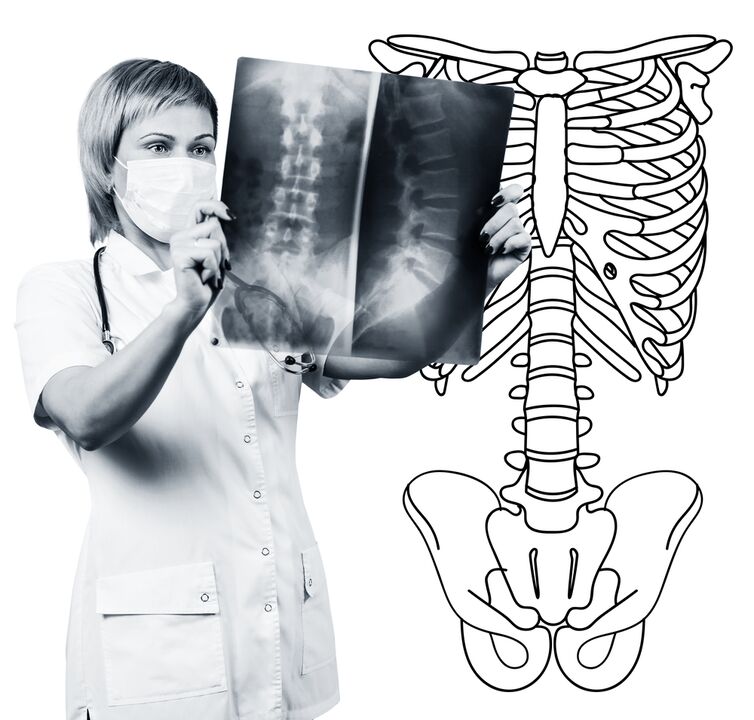
The diagnosis of lumbar spine osteochondrosis is based on clinical data and additional research methods.The main role belongs to methods such as:
- Radiography of lumbar spine;
- Computed tomography of lumbar spine;
- Magnetic resonance imaging of the lumbar spine.
Lumbar spine radiography is necessarily performed by 2 mutually exclusive forecasts-the rear and side.Such images allow you to see the shape, contours and structure of the spine bodies, the height and shape of the intervertebral discs, the spine pathologies and the natural curves.To show intervertebral joints and intervertebral holes, radiograms are produced in oblique projections.To determine the abnormal mobility of individual lumbar segments (which is a sign of osteochondrosis), radiography is performed in functional trial conditions, that is, in the flexibility and expansion of the spine.You can usually clearly see the height of the intervertebral discs in the front or back sections in the direction of the body prone to osteochondrosis, due to the functional block of one of the segments, the height of the disk does not change or bent or expand.Abnormal mobility is determined by the movement of the spine forward or back.The main X -ray signs of osteochondrosis include narrowing of the intervertebral wound, pathological movement and movement of vertebral bodies, deposit of salts (calcification) in disk tissue, regional growth of vertebral bodies, affected scerolosis (sub -labeling).Lumbar spine radiography is a routine of research that gradually loses its importance in the light of the active implementation of new and more informative research methods (CT and MRI).Radiography of the lumbar section is today used as a diagnostic method of screening.
The lumbar spine CT is also carried out using x -ray radiation, but the radial load on the body is much less than x -ray.The study is conducted on a special device table - a computed tomography, it is absolutely painless.The resulting images are processed using a computer and allow you to see significantly more structure than spine radiography.
MRI is a method in which electromagnetic radiation is used to create images.The study is also carried out on a table of deception on the table, which will fall into the tomography chamber.MRI is harmless and painless.
The lumbar spine CT or MRI allows you to view all the structures of the spine, carefully study the intervertebral discs (and the jacket and fiber rim) and intervertebral holes, the contents of the spinal canal.Even a slight protrusion of the intervertebral disc will not be overlooked.These methods (especially MRI) allow you to determine the disk hernia direction, if any, the degree of compression of the nerve roots, the spinal cord.Thus, these research methods are much more informative in the diagnosis of lumbar spine osteochondrosis than radiography.In addition, they allow you to diagnose not only osteochondrosis, but also other diseases (tumors, circulatory disorders in the spinal cord, abscesses, congenital defects of the spine and spinal cord structure), which are important in the background of the differential diagnosis of pain.
Lumbar spine osteochondrosis is a disease that most often causes pain.This is, in fact, the destruction of intervertebral discs.Due to osteochondrosis of the lumbar spine, a person often loses work capabilities because, in addition to pain, the disease can cause spine mobility, sitting, standing and walking.Symptoms of this disease are not specific and require additional research methods to accurately confirm the diagnosis.The most informative and safe of the modern methods of diagnosis of osteochondrosis diagnosis is the spine MRI.